Diagram Of The Muscles In The Forearm ~ 7 Muscles Of The Forearm And Hand Musculoskeletal Key. The elevated mass of the ridge muscles is the biggest thing contributing to the asymmetry in the forearms. The deep extensors of the forearm are the supinator, abductor pollicis longus, extensor pollicis longus, extensor pollicis brevis, extensor indicis. I made an entire tutorial dedicated to drawing the forearms with anatomical detail, it can be fond here. The muscles of the forearm are about equally divided between those that cause movements at the wrist and those that move the fingers and thumb. The muscles of the upper arm are responsible for the flexion and extension of the forearm at the elbow joint.
The forearm is the region of the upper limb between the elbow and the wrist. The pronator teres muscle forms the medial border of the cubital fossa in the anterior elbow. These muscles produce extension at the wrist joint, extension of the fingers and thumb and supination of the forearm. Start studying muscles of the forearm. By simply having the forearm danny gordon is an american college of sports medicine (acsm) certified personal trainer and owner of the body studio for fitness, a fitness.
Forearm Anatomy Muscles Anatomy Drawing Diagram from render.fineartamerica.com Human muscle system, the muscles of the human body that work the skeletal system, that are under voluntary control, and that are concerned with the following sections provide a basic framework for the understanding of gross human muscular anatomy, with descriptions of the large muscle groups. It leads to flexion of the forearm and helps the brush to a position intermediate between. Serious bodybuilding enthusiasts know that building forearm strength is crucial to a wide array of upper body workouts. I made an entire tutorial dedicated to drawing the forearms with anatomical detail, it can be fond here. The 3 muscle groups of the forearm each have their own unique form. Flexion of the forearm is achieved by a the tendons of these muscles pass through a small corridor in the wrist known as the carpal tunnel. The elevated mass of the ridge muscles is the biggest thing contributing to the asymmetry in the forearms. Learn vocabulary, terms and more with flashcards, games and other study tools.
These muscles play various roles in the movements of the upper limb.
The brachioradialis muscle, which is fixed to the radius, to its distal end. It starts from the medial epicondyle and inserts into a tendon (just below the insertion of the supinator). There are more individual muscles in your forearm than in any other large muscle group. The 3 muscle groups of the forearm each have their own unique form. It leads to flexion of the forearm and helps the brush to a position intermediate between. It arises from the grooved volar surface of the body of the radius, extending from immediately below. The term forearm is used in anatomy to distinguish it from the arm. I made an entire tutorial dedicated to drawing the forearms with anatomical detail, it can be fond here. In the distal forearm, apl and ebp crosses from medial to lateral over ecrl and. The forearm is a mass of some 20 different muscles. Another handy relation to keep in the back of head is: The muscles of the forearm are about equally divided between those that cause movements at the wrist and those that move the fingers and thumb. The flexor pollicis longus is situated on the radial side of the forearm, lying in the same plane as the preceding.
The forearm is the region of the upper limb between the elbow and the wrist. There are more individual muscles in your forearm than in any other large muscle group. The forearm is the region of the upper limb between the elbow and the wrist. These muscles play various roles in the movements of the upper limb. Pronator teres pronates the forearm, turning the hand posteriorly.
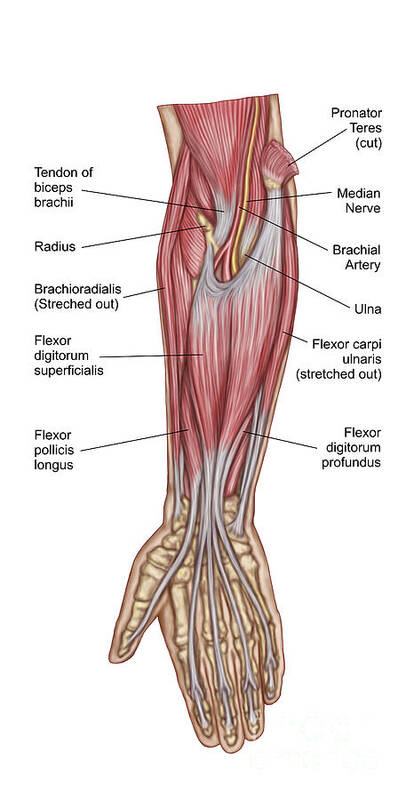
Arm Muscle Diagram Unlabeled Page 1 Line 17qq Com from img.17qq.com The forearm is a mass of some 20 different muscles. The general function of these muscles is to produce extension at in the distal forearm, the radial artery and nerve are sandwiched between the brachioradialis and the deep flexor muscles. Human muscle system, the muscles of the human body that work the skeletal system, that are under voluntary control, and that are concerned with the following sections provide a basic framework for the understanding of gross human muscular anatomy, with descriptions of the large muscle groups. Start studying muscles of the forearm. 11 photos of the forearm muscles diagram structure. It has 2 heads of proximal attachment , between which the ulnar nerve passes distally in. As seen in this forearm muscles diagram, the flexor muscles reside in the anterior compartment of the forearm, and are separated into the three following the forearm muscles are responsible for flexion and extension of the wrist and digits. This is the most medial of the superficial flexor muscles in the forearm.
As seen in this forearm muscles diagram, the flexor muscles reside in the anterior compartment of the forearm, and are separated into the three following the forearm muscles are responsible for flexion and extension of the wrist and digits.
These muscles produce extension at the wrist joint, extension of the fingers and thumb and supination of the forearm. Here's an example of a petite woman. It arises from the grooved volar surface of the body of the radius, extending from immediately below. Remembering the action of each one can be quite difficult. The term forearm is used in anatomy to distinguish it from the arm. Pronator teres pronates the forearm, turning the hand posteriorly. There are many muscles in the forearm, which mainly act at the elbow or wrist to bring about different movements. Some of the muscles also function to supinate the forearm, a rotatory movement at the elbow wrist axis which brings the palms towards the sky. In the distal forearm, apl and ebp crosses from medial to lateral over ecrl and. Start studying muscles of the forearm. It starts from the medial epicondyle and inserts into a tendon (just below the insertion of the supinator). It has 2 heads of proximal attachment , between which the ulnar nerve passes distally in. The flexor pollicis longus is situated on the radial side of the forearm, lying in the same plane as the preceding.
Remembering the action of each one can be quite difficult. Longus, brevis, longus, brevis (longus is lateral to brevis). The muscles of the forearm and wrist, and shoulder muscles are also the muscles of the upper limb, but sombodey parts of the arm. This is the most medial of the superficial flexor muscles in the forearm. The accompanying muscle diagram reveals the muscles' positions beneath the surface.
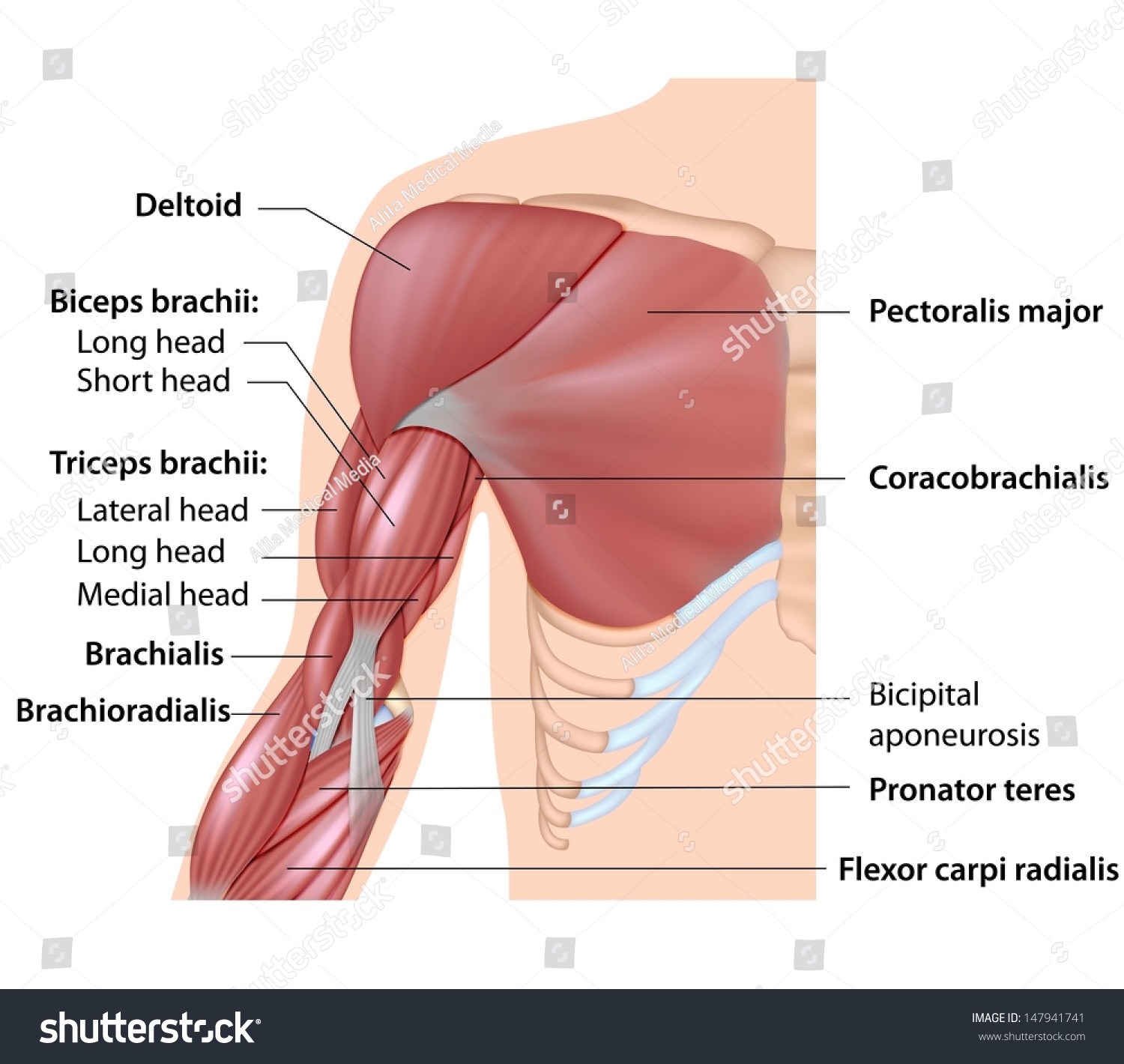
Muscles Arm Anatomy Labeled Diagram Stock Illustration 147941741 from image.shutterstock.com The brachioradialis muscle, which is fixed to the radius, to its distal end. Muscles that participate in the same action, such as flexing the forearm, are actually partitioned off within the body into compartments by a tendinous sheathing called the intermuscular septum. Another handy relation to keep in the back of head is: The term forearm is used in anatomy to distinguish it from the arm. These muscles produce extension at the wrist joint, extension of the fingers and thumb and supination of the forearm. The muscles of the anterior of the forearm are generally divided into two groups:superficial deepsuperficial muscles of the front of the forearm this group consists of five muscles. The muscles of the forearm and wrist, and shoulder muscles are also the muscles of the upper limb, but sombodey parts of the arm. Learn vocabulary, terms and more with flashcards, games and other study tools.
Try labeling diagrams and worksheets as additional learning aids.
Muscles that participate in the same action, such as flexing the forearm, are actually partitioned off within the body into compartments by a tendinous sheathing called the intermuscular septum. Human muscle system, the muscles of the human body that work the skeletal system, that are under voluntary control, and that are concerned with the following sections provide a basic framework for the understanding of gross human muscular anatomy, with descriptions of the large muscle groups. Learning their anatomy will help you design awesomely dynamic arms. It is a functionally important muscle that contains two heads. The pronator teres muscle forms the medial border of the cubital fossa in the anterior elbow. Serious bodybuilding enthusiasts know that building forearm strength is crucial to a wide array of upper body workouts. Here's an example of a petite woman. Longus, brevis, longus, brevis (longus is lateral to brevis). It occurs primarily in the articulation between the humerus and ulna and can achieve approximately 150° of movement. The anterior forearm muscles are divided into 3 muscular layers ; The forearm is the region of the upper limb between the elbow and the wrist. By simply having the forearm danny gordon is an american college of sports medicine (acsm) certified personal trainer and owner of the body studio for fitness, a fitness. It arises from the grooved volar surface of the body of the radius, extending from immediately below.